Posts

Showing posts from September, 2021

BURNS - RULE OF 9 IN BURNS,LATE COMPLICATION AND MANAGEMENT

Image
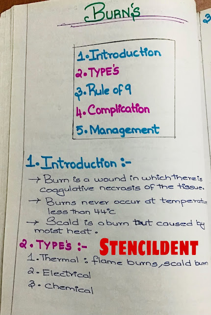
  BURNS BURN is a wound in which there is coagulative necrosis of the tissue  Burns never occur at temperature less than 44 degree celsius  Scald is a burn but caused by moist heat  TYPES: 1)THERMAL :-Flame burns,scald burn  2)ELECTRICAL 3)CHEMICAL RULE OF 9 IN BURNS : Pathology of burns are divided into : 1)Local changes: 2)Systemic changes  1)Local changes : Severity of burn Extent of burn Vascular changes Infection SEVERITY OF BURN: Microscopic destruction of the superficial layers of the epidermis Desquamated within few days  No scarring EXTENT OF BURN : Length and width of burn is expressed by percentage of total surface area displaying either 2nd or 3rd degree burn,extent estimated by WALLACE RULE OF NINES  ANATOMIC AREA                                               PERCENTAGE OF BODY SURFACE  Head ,face and neck                                                    9%  right upper extremity                                                 9% left upper extremity                     

BACTERIAL INFECTION - SYPHILIS ( ETIOLOGY,TYPES IN DETAIL NOTES )

Image
  SYPHILIS  SYPHILIS is a world wide chronic infection produced by treponema pallidum  History :christopher columbus and his crew returns from the new world possibly carrying syphilis Global epidemiology:15-49 years old- 6 million new cases each year Incidence decreased after penicillin Male is to female ratio is 1 is to 1 Fetal ,neonatal deaths- 3,00,000  ETIOLOGY: Treponema pallidum :gram positive ,motile ,spirochete and is pathogenic to human  ROUTE OF TRANSMISSION: Sexual contact  Vertical transmission  (mother to foetus) TYPES: 1)CONGENITAL  2)ACQUIRED : PRIMARY,SECONDARY,TERTIARY  1)CONGENITAL SYPHILIS : Transmitted to offspring only infected mother and not inherited  Totally preventable  Recognised if treatment with antibiotic is begun in infected pregnant women before 4 months of pregnancy  STIGMATA OF CONGENITAL SYPHILIS: 1)Head: Frontal bossing 2)Nose:Saddle nose 3)Eyes:Interstitial keratitis 4)Oropharynx;HUTCHINSON'S TRIAD  HYPOPLASIA OF INCISOR -screw driver shaped inci

SEBACEOUS CYST- CLINICAL FEATURES,TREATMENT,COMPLICATION

Image
  SEBACEOUS CYST  INTRODUCTION: SYNONYMS: EPIDERMOID CYST ,WEN  Etiology: acquired -retention cyst ,sebaceous cyst -obstruction  leads to accumulation of sebaceous material  Site:  Face Scalp Scrotum Back It does not occur in palm and sole cause sebaceous glands are absent  CLINICAL FEATURES: AGE:Early adulthood,middle age  Slow growing Shape:Hemispherical,Spherical  Central keratin filled- punctum indicate blockage of duct ,dark spot  20-30% cases punctum may not be seen  Surface :smooth,round border  Consistency :soft and putty,non-tender Sign of moulding Sign of indentation :pitting on pressure over the swelling Swelling mobile,except at the site of punctum Bony defect -absent Multiple sebaceous cyst may be associated with gardner syndrome TREATMENT: Incision and avulsion of cyst with the wall During dissection,cyst wall ruptures ,care should be taken to excise cyst wall ,cause there can be recurrence Small -excised with skin COMPLICATION: INFECTION: Injury,scratch may lead  to absc

PERITONSILLAR ABSCESS -ETIOLOGY,MECHANISM,CLINICAL FEATURES,TREATMENT

Image
  PERITONSILLAR ABSCESS ( QUINSY) 1)INTRODUCTION: TONSIL-Subepithelial aggregation of lymphoid tissue which forms a part of waldeyer's ring Its ovoid in shape Situated in lateral wall of oropharynx QUINSY- Collection of pus in peritonsillar space between capsule and superior constrictor muscle 2)ETIOLOGY: As a sequelae of acute tonsillitis De novo  Causative organism :  Streptococcus pyogenes Staphylococcus aureus  Anaerobic organism   3)MECHANISM : One of the crypts or crypta magna gets filled 

CHERUBISM /FAMILIAL FIBROUS DYSPLASIA OF JAWS -Definition,Pathogenesis,Clinical features,Radiographic features,Histopathology and Treatment

Image
  CHERUBISM SYNONYMS: 1)FAMILIAL FIBROUS DYSPLASIA OF JAWS 2)DISSEMINATED JUVENILE FIBROUS DYSPLASIA  INTRODUCTION: JONES in 1933  coined the term cherubism  They resemble cherubs ( chubby cheeked little angel in renaissance painting) Autosomal dominant fibro-osseous lesion of jaws DEFINITION : Rare inherited autosomal dominant disease that causes bilateral enlargement of jaws giving the child a cherubic facial appearance Regress with age ,composed of giant cell granuloma like tissue and does not form bone matrix  PATHOGENESIS: Mutation in gene coding SH3BP2 located on 4P16.3  Autosomal dominant trait  Deficiency of sex steroids leads to reduction in osteoclast formation which leads to excess bone formation  At puberty, Osteoradiol+testosterone will cause increase in plasma concentration which results in localised increase in osteoclast  Thereby,stabilizes the disease  CLINICAL  FEATURES: Male's are more commonly affected than female ,100%penetrance in males,50-70% in females Age:

Technique To Strengthen Ceramic

Image
  STRENGTHENING CERAMIC   Ceramic brittle matter and have low tensile strength - this makes it chip of at certain clinical condition. Therefore necessary to strengthen ceramic to increase longevity.  TECHNIQUE : 1)MINIMISING THE EFFECT OF STRESS CONCENTRATION: A)DESIGN OF PROSTHESIS: Select stronger and tougher ceramic  Grinding of surface of ceramic restoration should be minimised Avoid sharp line angle in tooth preparation  2)DEVELOPMENT OF RESIDUAL COMPRESSIVE STRESS: In post metal ceramic restoration : Metal should have more thermal coefficient of expansion than ceramic  Induce compressive stress this can withstand fracture  In all -ceramic : core ceramic have more thermal coefficient of expansion  3)MINIMISING NUMBER OF FIRING CYCLES: Repeated firing will result in : weaker and fragile ceramic If we reduce the number the number of firing we can strengthen ceramic  4)THERMAL TEMPERING : Ceramic is heated to a higher temperature ,once it reaches high temperature its quenched rapidly