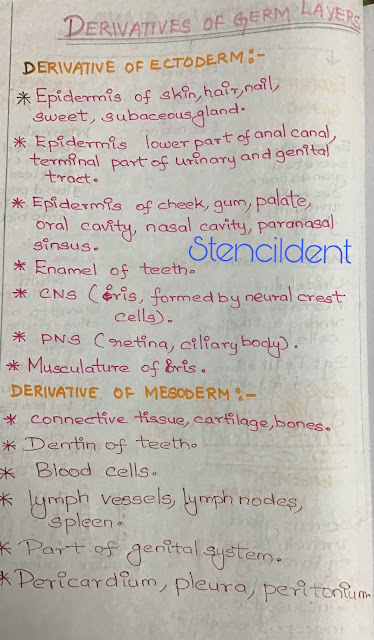
Posts

Showing posts with the label ANATOMY NOTES

Cavity of larynx anatomy

Image
 CAVITY OF LARYNX Larynx extent: inlet of larynx to the cricoid cartilage Relation:  ANTERIOR:  epiglottis POSTERIOR; Interarytenoid fold Internal diameter : 3 mm upto 3 years and increases 1mm upto 12 years . Within cavity of larynx two folds are present .upper fold,vestibular and lower fold,vocal The space between vestibular fold is rima vestibule,vocal fold is rima glottis . Vocal fold is limited anterior by interarytenoid fold  Rima is narrowest portion of larynx 23 mm in males and 17mm in female    HANDWRITTEN NOTES ON CAVITY OF LARYNX  REFER GOOGLE DIAGRAM TO SEE REAL IMAGE OF LARYNX  Vestibular fold and vocal fold is dividing the cavity of larynx into three : Region above vestibular fold: vestibule,  supraglottis. Region between vestibular fold and vocal fold: ventricle Region below vocal fold: infraglotis Sinus of morgagni lies between vestibular and vocal fold. Sinus of morgagni /ventricle of larynx extend anteriorly till the lamina of thyroid ,cartilage is known as saccule of

Derivatives of germ layers

Image
  DERIVATIVE OF GERM LAYERS INTRODUCTION : 1)Germ layers arise from a process of gastrulation ,an early phase of embryological development in which implanted blastula develops into 3 germ cell layers ,occur during 3rd week of development. 2) Its marked by extensive cell movement where blastula  becomes organised into 3 layers .  MORULA ( a ball of cells) ,BLASTULA(hallow ball) ,gastrulation ( 3 layers of hollow ball ) 3) BLASTOCOEL: fluid filled cavity that forms in the blastula of early amphibian and its functions is to permit cell migration during gastrulation and to prevent cell beneath it from interacting prematurely with other cells . DERIVATIVE OF ECTODERM : 1) Epidermis of skin ,hair,nail,sweat,sebaceous gland . 2) epidermis lower part of anal canal,terminal part of urinary and genital tract . 3)epidermis of cheek,gum,palate,oral cavity,nasal cavity,para nasal sinus 4) enamel of teeth  5) central nervous system : formed by neural crest cells  6) peripheral nervous system : retin

VERTEBRAL ARTERY

Image
    VERTEBRAL ARTERY  Vertebral artery is principal artery that supplies  brain,spinal cord,meninges and i t has 4 parts.

NERVE SUPPLY OF SCALP - LEARN IT EASY WITH STENCILDENT

Image
              " NERVE SUPPLY OF SCALP"    INTRODUCTION 10 nerves supply scalp ,8 nerve via sensory root and 2 nerve via motar root . sensory nerves pass further behind the auricle and in front of auricle       Its one 5 mark question which came in our university paper so get prepared well u might also get it this question.                         THANKS FOR WATCHING 

ARTERIAL SUPPLY OF SCALP - LEARN IT EASY STENCILDENT

Image
                               " ARTERIAL SUPPLY                                OF SCALP" Scalp is a soft tissue covering the cranial vault.         IN FRONT OF AURICLE          supratrochlear         supraorbital         superficial temporal artery          BEHIND AURICLE         POSTERIOR AURICULAR         OCCIPITAL ARTERY         THANKS FOR WATCHING 

CAROTID SHEATH - LEARN IT EASY WITH STENCILDENT

Image
                      "  CAROTID SHEATH " Condensation of fibro areolar tissue around main vessel of neck. ITS formed anterior aspect -pretracheal fascia , posterior aspect-prevertebral fascia                      THANKS FOR WATCHING 

ANSA CERVICALIS : ANATOMY NOTES LEARN IT EASY WITH STENCILDENT

Image
                      ANSA CERVICALIS  Thin nerve loop lies embedded  in anterior wall of carotid sheath in lower part of larynx. It supplies infrahyoid muscle  hope this would have been useful  THANKS FOR WATCHING 

DIGASTRIC TRIANGLE

Image
                DIGASTRIC TRIANGLE     INTRODUCTION  Its also know as submandibular triangle,paired triangle in anterior region of neck . NOTE: REFERENCES :B.D.CHAURASIA 

STERNOCLEIDOMASTOID MUSCLE

Image
                       STERNOCLEIDOMASTOID                           MUSCLE        Its enclosed in the investing layer of deep cervical fascia pierced by accessory nerve. thanks for watching 

DEVELOPMENT OF FACE

Image
THE DEVELOPMENT OF FACE  The face develops from 5 swellings that form around the primitive mouth (stomodeum). single  fronotonasal process  paired maxillary process  paired mandibular process  FRONTONASAL  processes forms:    forehead ,external nose,nasal cavity,nasal septum and philtrum of lip.   MAXILLARY process forms : lateral parts of upper lip and upper part of cheek MANDIBULAR process forms : lower lip and lower part of cheek . CLINICAL ANATOMY : CLEFT LIP AND CLEFT PALATE