Posts

Showing posts with the label Pathology

Dystropic calcification :Pathogenesis,morpholgy ,histology

Image
 DYSTROPHIC CALCIFICATION  Pathologic calcification is abnormal tissue deposition of calcium salts together with smaller amounts of iron ,magnesium and other mineral salts Its of two forms : Dystrophic Metastatic Dystrophic calcification : When deposition occurs locally in dying tissue It occurs despite normal serum level of calcium and in the absence of derangement in calcium metabolism Occur in : Area of necrosis Advanced atherosclerosis Damaged heart valve Dead parasite Cancer Students corner : To learn more about  pathogenesis of atherosclerosis  do click on this link to learn more. Pathogenesis: Final step if fomrtaion of crystalline calcium phosphate  INITIATION: Membrane process calcium concentration bind to phospholipid present in membrane ,phosphatase generate phosphate group   PROPAGATION: Cycle of calcium binding phosphate generating micro crystal propagate lead to more calcium deposition  MORPHOLOGY : Calcium salts appear macroscopically fine ,white granules or clum ,gritt

Disseminated intravascular coagulation- MIND MAP

Image
    DISSEMINATED INTRAVASCULAR                                          COAGULATION INTRODUCTION: Complex thrombohemorrhagic disorder Second complication in some disease ETIOLOGY: Massive trauma,surgery Malaria Septicemia Severe burn Snake bite  PATHOGENESIS: Activation of coagulation Thromboitic phase Consumption phase Secondary fibrinolysis CLINICAL FEATURE : Bleeding Organ damage LABORATORY DIAGNOSIS: Platelet count low  Prothrombin and thrombin time is prolonged

Pathogenesis behind atherosclerosis

Image
  PATHOGENESIS OF ATHEROSCLEROSIS INTRODUCTION: Lipid plaque form on the inside wall of arteries Arteries :narrowed and becomes hardened Atherosclerosis is caused when endothelial wall get damaged by hypertension ,smoking,hyperglycemia Increased permeability of arterial wall -allowing LDL cholesterol to get in  White blood cells like monocyte ,move freely through blood vessel Endothelial cell express adhesion protein molecule when an irritating stimuli ,tat capture monocyte ,undergo morphologic changes ,enter between cell -DIAPEDIESIS White blood cell (engulf )produce free radical in contact with LDL  gets oxidized resulting in positive feedback situation  Foam cell produced ,saturated with LDL die and release content ,that's taken by other white blood cell Fragment of dead cell produce area with lipid core to form plaque This plaque accumulate more calcium salt and dead cell leading to atherosclerosis Blood clot form on endothelial wall thrombus Flow through blood stream called em

VIRCHOWS TRIAD-thrombus formation

Image
  VIRCHOWS TRIAD  THROMBOSIS : Process of formation of solid mass in circulation from flowing blood ,this mass itself is called thrombus According to virchows three things/factors essential for blood clot ,"thrombus formation". 1)Endothelial cell injury 2)Hemostasis 3)Hypercoagulality STUDENTS CORNER : Welcome back to our site ,if you are our new subscriber then do consider checking out our  posts by either clicking on the subject you want to learn or you can start by searching the topic of your interest ,coming back to the content so today the topic of discussion is on virchows triad ,previously we have discussed on  PLATELET , FATE OF THROMBI , when this question is asked do give a short introduction on platelet and proceed by mentioning all three in the virchows triad that is the endothelial injury,hemostasis( click on the link to learn about hemostasis ) followed by hypercoaguability and conclude your answer with fate of thrombi . ENDOTHELIAL INJURY: Increases the risk

PACKED CELL VOLUME ?USES,METHODS OF COLLECTION,ITS SIGNIFICANCE AND REFERENCE RANGES

Image
                       PACKED CELL VOLUME   Its also called hematocrit Volume occupied by red cell when sample of anticoagulated blood is centrifuged It indicates relative proportion of red cell to plasma It is expressed as percentage of original volume of blood or as decimal fraction USES: Detection of presence /absence of anemia or polycythemia Estimation of red cell indices (mean cell volume +mean corpuscular Hb concentration) Checking accuracy of Hb value (Hb in g/dl *3=PCV) METHODS: Two methods for estimation Macro(WINTROBE),MICRO(MICROHEMATOCRIT) Micro method is preferred : Rapid Convenient Small amount of blood After centrifugation of anticoagulated three zones can be seen above downward: Plasma Buffy coat Packed cell Smear from puffy coat for demonstration of lupus erythematosus, malaria parasite and immature cells plasma IS usually Straw coloured  If colorless-IRON DEFICIENCY PINK-HEMOGLOBINEMIA YELLOW-JAUNDICE MILKY-HYPERTRIGLYCERIDEMIA SIGNIFICANCE: In anemia :Packed cell vo

AGRANULOCYTOSIS -Causes, symptoms ,risk ,diagnosis and treatment

Image
                   AGRANULOCYTOSIS Lack of granulocyte  Absolute neutrophil count (less than 100 per microliter) Rare condition Suppressed immune system CAUSES: May be inherited or genetic Drugs: Anti-thyroid ,Anti-psychotic ,antibiotic Exposure to toxic substance such as ARSENIC ,MERCURY Aplastic anemia SYMPTOM : Sore throat and mouth Fever ,chill Fatigue  Headache Sweating  Swollen gland RISK: If left untreated result in sepsis Male and female both at equal risk Certain drug  DIAGNOSIS: Blood test Neutrophil count less than 100 per microliter of blood Bone marrow biopsy TREATMENT: Change of drug Transfusion of granulocyte 

MUST KNOW FACTS ABOUT CYTOKINES

Image
                                     CYTOKINES  INTRODUCTION: Cells communicate at molecular level by cytokine Paracrine function: released peptide and other molecule Soluble protein SECRETD BY : Hemopoietic  Non-hemopoietic cells in response to various stimuli ROLE: Activaton of immune system  PROPERTIES EXHIBITED: PLEIOTROPHIC: One cytokine act on different cell type REDUNDANCY: Similar function by different cytokine Cascade induction: one cytokine stimulate its target cell to make another cytokine SYNERGY: Combination cytokine result in combined effect  CATEGORIES: INTERFERON-involved in antiviral response  INTERLEUKIN-produced by one leucocyte, act on other TUMOUR NECROSIS FACTOR TRANSFORMING GROWTH FACTOR COLONY STIMULATING FACTOR-support growth of blood cell GROWTH FACTOR Growth factor and other cytokine -CRINOPECTIN Cytokine involved in leukocyte endothelial cell interaction -CHEMOKINE  INTERLEUKIN: About 35 interleukin been identified  Example : T-Cell secrete -INTERLEUKIN -2 a

DAY IN MY LIFE OF THROMBUS /FATE OF THROMBUS?

Image
                             FATE OF THROMBI Lets  have a sneak peak of how thrombi(blood clot) usually spends its precious time in our body. Either of these 4 jobs is what thrombi usually does : RESOLUTION,ORGANISATION ,PROPOGATION AND THROMBOEMBOLISM . STUDENTS CORNER: Before getting into the topic we must understand a few terminologies to get a picture of how thrombus spends its day: firstly what is Thrombosis's a process of formation of solid mass in circulation from flowing blood . Mass itself is known  as thrombus. Progression of thrombus is what is meant by fate of thrombus. If the question is about thrombosis then we need to give short introduction of THROMBOCYTE  ,thrombus, thrombosis etiopathogenesis, Virchow's triad,  and then conclude with the fate of thrombus. RESOLUTION/DISSOLUTION: Thrombus activate fibrinolytic system to release plasmin ,this plasmin dissolve the thrombus which results in RESOLUTION  Small venous thrombi undergoes complete lysis Large thrombi ma

NECROSIS -pathology notes

Image
  NECROSIS  DEFINITION: Its defined as the morphological changes taking place in a tissue after cell death due to degradative action f enzymes on irreversibly injured cell. TYPES OF NECROIS : A)CASEOUS NECROSIS B)LIQUEFACTIVE NECROSIS C)COAGULATIVE NECROSIS D)FIBRINIOD NECROSIS E)FAT NECROSIS F)GANGEROUS NECROSIS CYTOPLASMIC CHANGES : Increased eosinophilia of cytoplasm  Vacuolation and moth eaten appearance of cytoplasm  Calcification of cell membrane and intracellular structures  Formation of myelin figures  NUCLEAR CHANGES : KARYOLYSIS : Fading of basophilia of nucleus PYKNOSIS: Nuclear shrinkage due to condensation of chromatin  KARYORRHEXIS -Nuclear fragmentation  A)CASESOUS NECROSIS : Its seen in tuberculosis and occurs due to both enzymatic degradation of tissue and denaturation of cellular proteins  The tissue architecture is lost and replaced by soft ,creamy cheese like material  Microscopically ,there is amorphous granular debris seen  Examples : tuberculosis lymph node ,tube

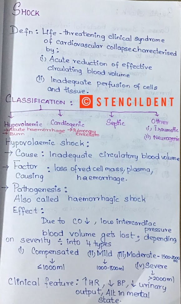
Shock pathology notes

Image
                               SHOCK  HELLO! DOCTOR VANAKAM,  The topic of discussion for today is SHOCK as I had already mentioned it in my previous post on sterilization techniques in microbiology if u haven't read it so far check it out using this link  https://www.stencildent.com/2020/07/sterilization-technique-in-microbiology.html  without any further ado lets get started. CONTENTS: 1) Definition 2) Classification 3)Hypovolaemic shock- pathogenesis 4)Septic shock 5)Etiology and pathogenesis of circulatory shock 6) Complication  1) DEFINITION : It's a life-threatening clinical syndrome of cardiovascular collapse characterized by acute reduction of effective circulating blood volume, inadequate perfusion of cells and tissue. 2) CLASSIFICATION : Shock is classified into hypovolaemic, cardiogenic, septic, other shocks which include traumatic and neurogenic 3) HYPOVOLAEMIC SHOCK: Cause:  inadequate circulatory blood volume  The factor causing: loss of red cell mass, plasma, he